Home

casquette Nord coupon blood clot in leg ultrasound Impoli Morbidité Appartement

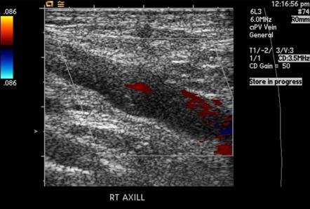
Upper Extremity DVT | Cedars-Sinai

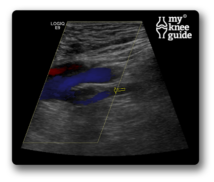
Phlebitis | The VeinCare Centre

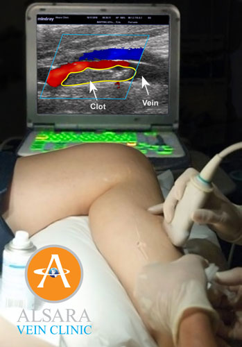
Blood Clots & Varicose Veins - 5X Higher Risk - Alsara Vein Clinic

Ultrasonography of deep vein thrombosis - Wikipedia

Blood Clots: Also known as a DVT

Deep vein thrombosis | Radiology Reference Article | Radiopaedia.org

Vein-Venous Doppler Ultrasound | pictureperfectultrasound

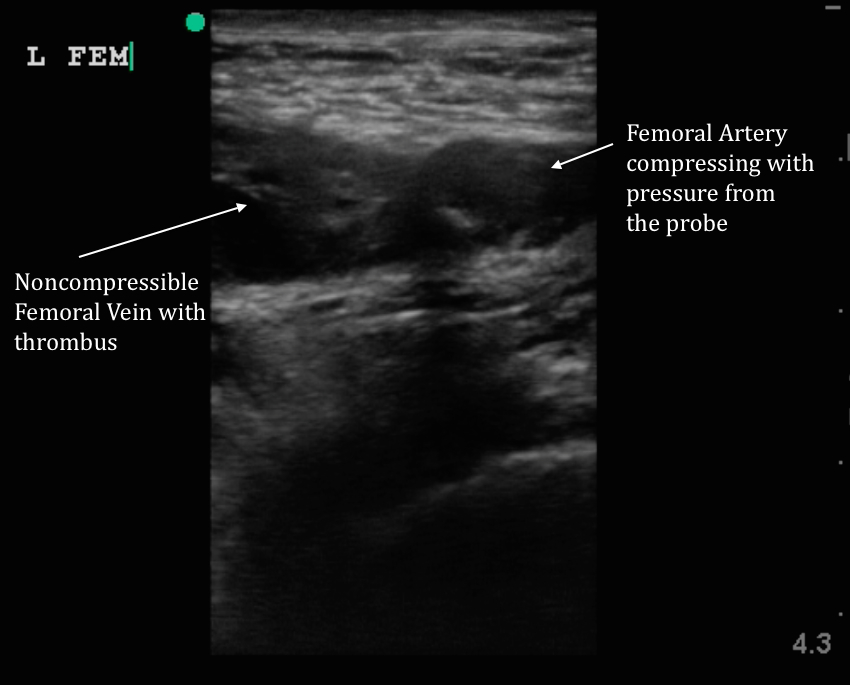
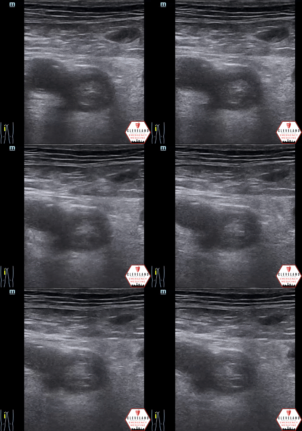
Intern Ultrasound of the Month: DVT Diagnosed at the Bedside — University  Hospitals Emergency Medicine Residency
Intern Ultrasound of the Month: DVT Diagnosed at the Bedside — University Hospitals Emergency Medicine Residency
Deep Vein Thrombosis Bedside Ultrasonography: Practice Essentials,  Preparation, Technique
Deep Vein Thrombosis Bedside Ultrasonography: Practice Essentials, Preparation, Technique

DVT Evidence — TPA
DVT Evidence — TPA

Massive DVT | Emory School of Medicine

Residual Clot: What is it? Should it affect management?

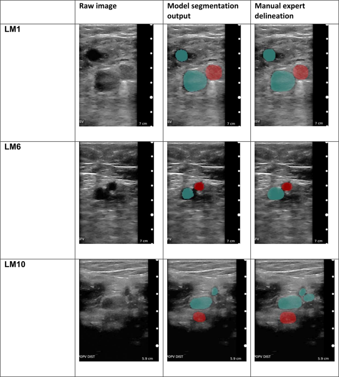
Non-invasive diagnosis of deep vein thrombosis from ultrasound imaging with  machine learning | npj Digital Medicine
Non-invasive diagnosis of deep vein thrombosis from ultrasound imaging with machine learning | npj Digital Medicine

Deep vein thrombosis - Wikipedia

Chronic recanalized deep vein thrombosis on ultrasound. Gray scale... |  Download Scientific Diagram
Chronic recanalized deep vein thrombosis on ultrasound. Gray scale... | Download Scientific Diagram

DVT ultrasound protocol. Step by step guide to ruling out a DVT

Doppler ultrasound showing blockage of the left superficial femoral... |  Download Scientific Diagram
Doppler ultrasound showing blockage of the left superficial femoral... | Download Scientific Diagram

Venous Ultrasound: VU - SONOSIF - by SIFSOF, California

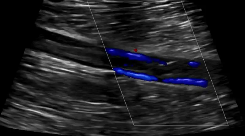
Doppler ultrasound showing deep vein thrombosis - Stock Image - M175/0277 -  Science Photo Library
Doppler ultrasound showing deep vein thrombosis - Stock Image - M175/0277 - Science Photo Library

Deep vein thrombosis - Harvard Health

5 Signs of a Blood Clot: Dallas Vein Institute: Varicose Vein Specialists

A-D. Clinical condition of deep vein thrombosis. A) A poor blood flow... |  Download Scientific Diagram
A-D. Clinical condition of deep vein thrombosis. A) A poor blood flow... | Download Scientific Diagram
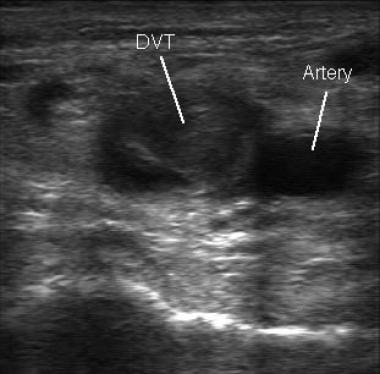
Ultrasound Leadership Academy: Lower Extremity DVT — EM Curious

Ultrasound for Lower Extremity Deep Venous Thrombosis | Circulation

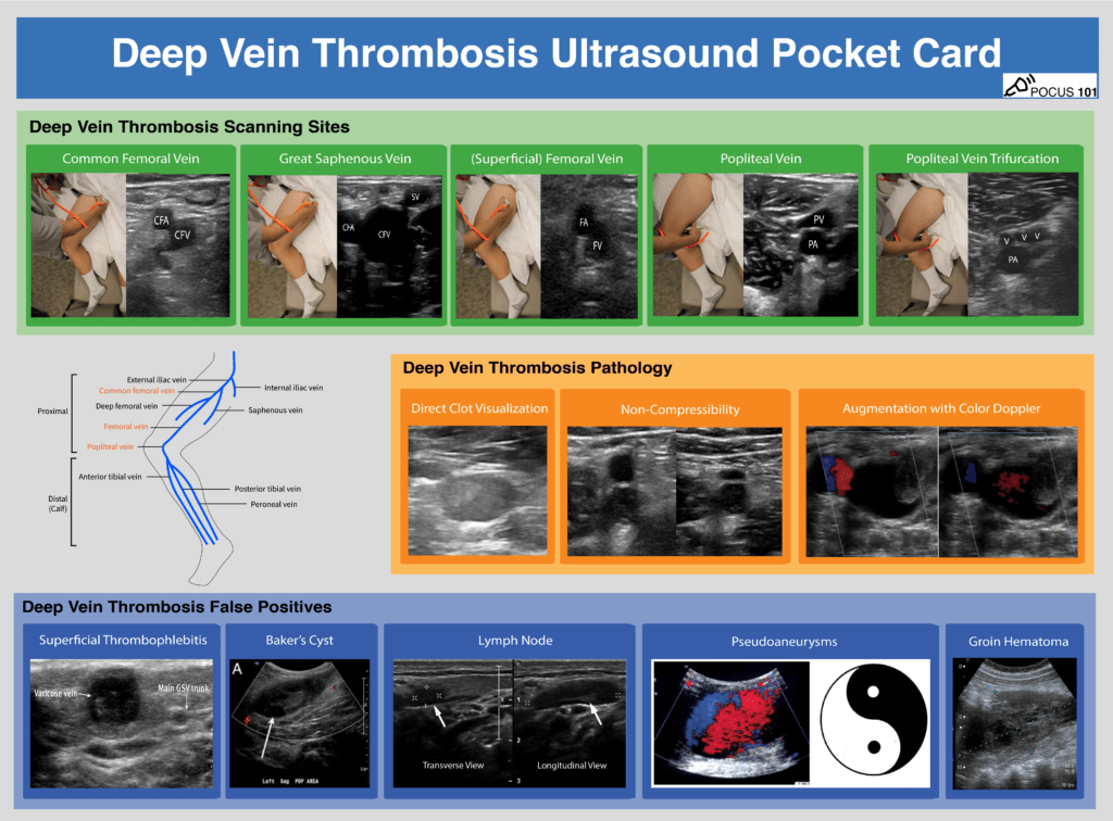
DVT Ultrasound Made Easy: Step-By-Step Guide - POCUS 101

Blood Clots in Legs: Causes, Diagnosis and Treatment